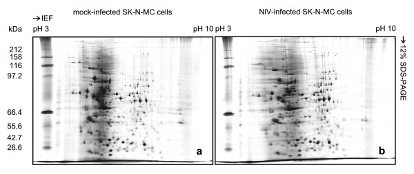
Figure 1.
Two-dimensional gel electrophoresis of mock-infected and NiV-infected SK-N-MC cells. Mock-infected and NiV-infected cell proteins were extracted directly using urea buffer. IEF was performed in 7 cm IPG strips, pH 3–10 using 100 μg of mock-infected (a) and NiV-infected (b) SK-N-MC cell proteins.

