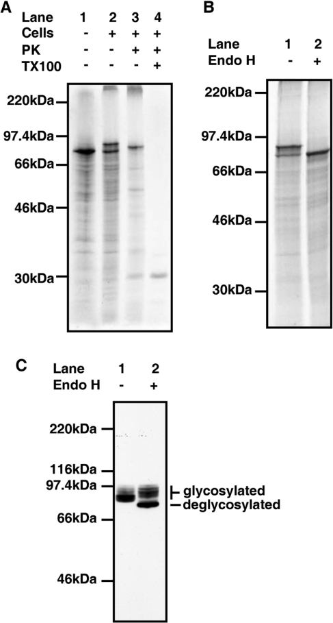
Figure 1. In vitro translations and in vivo processing of hQSOX1a.
(A) hQSOX1a-V5 mRNA was translated in a rabbit reticulocyte lysate in the absence (lane 1) or presence of SP HT1080 cells (lanes 2–5) for 1 h at 30°C. Isolated cells were treated with Proteinase K (PK; lane 3) with the addition of 1% (w/v) Triton X-100 (TX100) as indicated (lane 4). (B) hQSOX1a-V5 mRNA was translated in a rabbit reticulocyte lysate in the presence of SP HT1080 cells for 1 h at 30°C. Isolated cells were incubated with (lane 2) or without (lane 1) Endo H at 37°C for 1 h. (C) Cells stably expressing hQSOX1a-V5 were lysed and proteins were either treated (lane 1) or not treated (lane 2) with Endo H. Proteins were detected by immunoblotting with V5-antibody.

