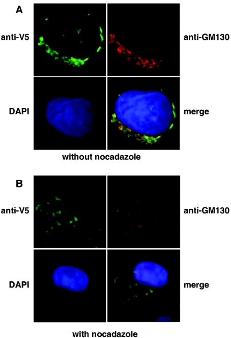
Figure 2. Intracellular localization of V5-tagged hQSOX1a.
(A) hQSOX1a-V5 expressing CHO-tPA cells were fixed and simultaneously stained with mouse anti-V5 antibody and rabbit anti-GM130 antibody. Secondary antibodies were conjugated to Alexa Fluor® 448 (green) or Alexa Fluor® 594 (red). Cell nuclei were stained with DAPI. (B) Golgi localization of the hQSOX1a-V5 was confirmed by incubating cells in the presence of 5 μg/ml nocadazole for 2 h. Cells were then fixed and stained as above.

