
Figure 1.
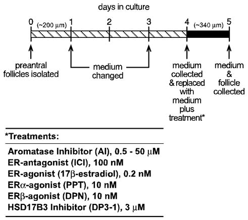
Scheme used for in vitro culture of wild type and αERKO follicles for the assessment of steroidogenesis and gene expression. Large preantral follicles of approximately 200 μm in diameter were isolated from immature (21–24 d) wild type and αERKO females and individually propagated in a 250 μl volume of medium for 5 days, during which they grow to approximately 340 μm in diameter. Media was changed and or collected on the indicated days. Follicles were inspected for integrity and health every 24 h and removed from the study if they did not satisfy the conditions described in the Materials and Methods. The 24 h period between days 4–5 was found to be the period of peak steroid synthesis by follicles and was therefore selected for all experimental treatments. Media was changed on day 4 of culture and replaced with media containing vehicle or a combination of the indicated treatments. The media was then collected after 24 h and stored for evaluation of androstenedione, testosterone and estradiol content by EIA. The follicle was also collected for later evaluation of gene expression.