
Figure 6.
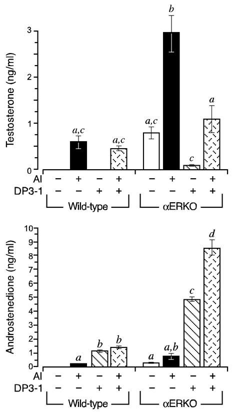
Ectopic HSD17B3 activity accounts for the aberrantly elevated capacity for testosterone synthesis in αERKO follicles. Wild type and αERKO follicles were grown as described in Fig. 1. During the 24 h culture period between days 4–5, follicles were exposed to an aromatase inhibitor (AI) and/or an HSD17B3-specific inhibitor (DP3-1). Shown is the average (± SEM) amount of testosterone (top) and androstenedione (bottom) synthesized during the 24 h period. As was first shown in Fig. 2, both wild type and αERKO follicles synthesized increased amounts of androstenedione and testosterone when exposed to an AI, however, the levels of both androgens are much higher in the αERKO follicles. Furthermore, testosterone synthesis in αERKO follicles is inhibited by DP3-1, indicating it is mediated by HSD17B3, a Leydig cell-specific enzyme. In contrast, DP3-1 had no effect on testosterone synthesis in wild type follicles, indicated this is likely mediated by the androgenic properties of rodent HSD17B1. The data shown is one of two independent experiments that yielded comparable results. Sample sizes were 8–9 follicles per genotype, per treatment, per experiment.