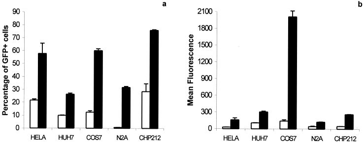
Figure 1.
Expression of the GFP gene in various mammalian cells infected with Bac-CMV-GFP. 105 cells were infected with Bac-CMV-GFP at a moi of 10 and harvested 24 h after infection. Three wells were infected for each condition. In the condition of induction by sodium butyrate (5 mM), the deacetylase inhibitor was added to the medium just after the virus was removed. (a) Percentage of cells expressing GFP, expressed as the average percentage of cells that were GFP+ in three independent transductions ± SD. (b) Average intensity of fluorescence for a transduced cell, expressed as the mean of fluorescence of three independent transductions. Empty columns represent infected cells, not treated with sodium butyrate. Filled columns represent infected cells treated with 5 mM sodium butyrate.

