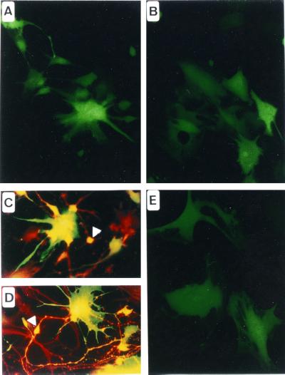
Figure 3.
Transduction of human primary neural cultures with Bac-CMV-GFP. (A) Transduction of embryonic telencephalic cells cultured in presence of basic fibroblast growth factor. Morphologically, transduced cells appear to be of neuroepithelial and neuronal phenotypes. (B) Transduction of embryonic telencephalic cells cultured in presence of FCS. Morphologically, cells appear to be of glial lineage. (C) Anti-Map2 immunochemistry. The arrowhead shows a transduced neuron. (D) Anti-β3-tubulin immunochemistry. The arrowhead indicates a transduced neuroepithelial cell. (E) Transduction of primary human adult astrocytes.

