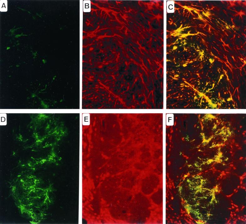
Figure 5.
Histological analysis of in vivo gene injection of Bac-CMV-GFP into the striatum of BALB/c adult mouse, 48 h after delivery. (A and D) Anti-GFP immunochemistry. (B) Anti-GFAP immunochemistry. (C) GFP and GFAP double labeling. Note that most of the cells are astrocytes. (E) NeuN immunochemistry. (F) GFP and NeuN double labeling. Note that very few transduced cells are of neuronal phenotype.

