Figure 4.
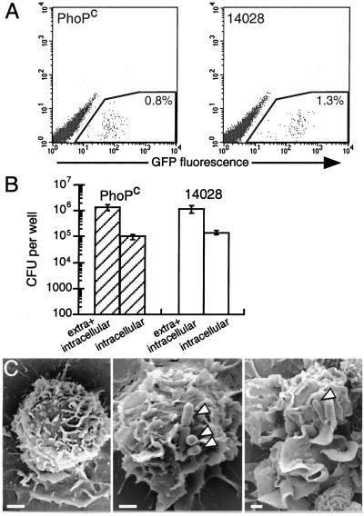
Entry of S. typhimurium in DCs. (A) Adhesion of S. typhimurium to DCs. S. typhimurium PhoPc and wild-type expressing GFP were incubated at 4°C for 30 min with DCs. After washing, the cell suspension was analyzed by flow cytometry. The proportion of cells associated with the fluorescent bacteria is indicated in the gated region. The figure shows a representative experiment of three independent experiments performed. (B) Internalization of S. typhimurium in DCs. S. typhimurium PhoPc (hatched bars) and wild-type (open bars) strains were incubated for 30 min with DCs. After washing, the cells were lysed and plated onto agar to count the number of extracellular and internalized bacteria, or incubated for 30 min with gentamicin to kill extracellular bacteria, lysed, and plated onto agar to count the internalized bacteria. The figure shows the mean ± SD of four independent experiments performed in duplicate. (C) Scanning electron micrographs of DCs not treated (Left) or incubated for 3 min with S. typhimurium PhoPc (Middle), or wild-type (Right). Arrowheads point to bacteria. Bar = 1 μm.

