Figure 3.
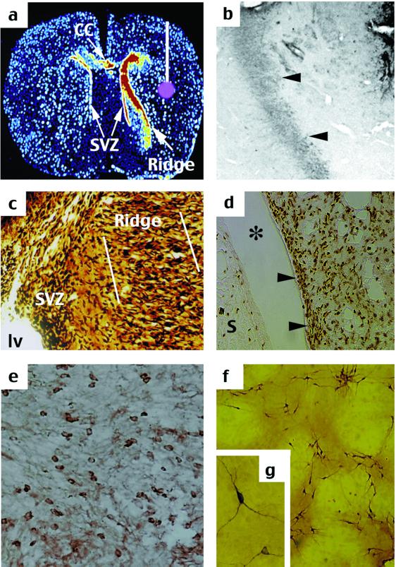
Further characterization of the TGFα-induced striatal ridge cells. (a) Cross section of pseudocolor autoradiographic image (EGFr-mRNA) showing SVZ, location of a ridge, a smaller ridge in the corpus callosum (CC), and a cartoon image of the TGFα infusion cannula (white line) and infusion site in the right caudate putamen (pink circle). (b) The ridge cells (arrows) are nestin-positive, showing that they are neural progenitors. (c) Silver staining shows a fusiform morphology of the cells in the ridge (outlined by white lines), suggestive of outward migration from the SVZ lining the lateral ventricle (lv). (d) BrdUrd was incorporated by SVZ (arrows) and ridge cells laterally in the striatum, but not in the septum (S) after a striatal TGFα infusion. (e) Some migrating cells subsequently stained positive for β-III tubulin, a marker for neuronal restricted lineage. (f) Longer TGFα infusion times revealed increasing numbers of TH-positive neurons (higher magnification in g).

