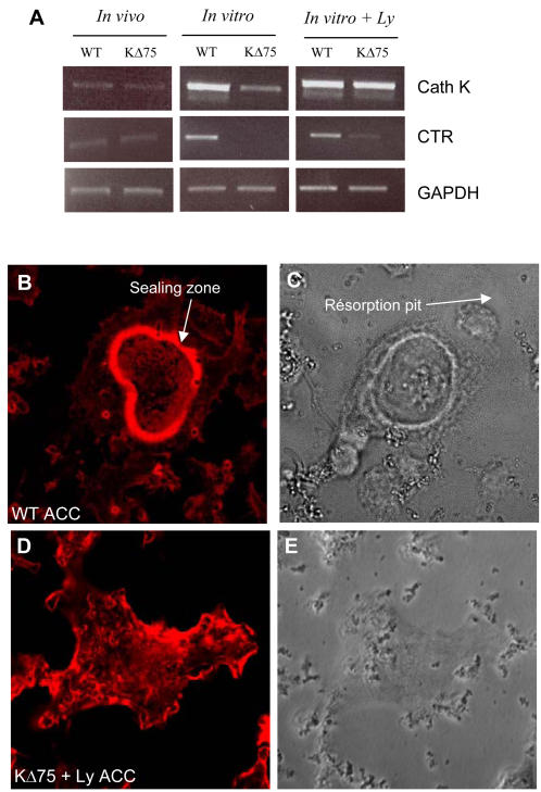
Figure 3. Lymphocytes trigger osteoclast marker expression but not bone resorption from KΔ75 hematopoietic precursors.
Expression of cathepsin K (Cath K) and calcitonin (CTR) receptors by RT-PCR. RNAs were extracted from WT and KΔ75 mouse spleen cell cultures (in vitro) maintained for 7 days with MCSF and RANKL, with or without (+Ly) lymphocytes. As a control, RNAs from WT and KΔ75 mouse bones (in vivo) were also used. Under in vivo conditions, both Cath K and CTR were expressed in WT and KΔ75 mouse bones, as well as in in vitro cultures from corresponding spleen cell cultures. However, in the absence of lymphocytes in in vitro cultures, Cath K is weakly expressed and CTR is absent from KΔ75 mouse cell cultures. (B–E) Functional resorption tests on mineralized ACC matrix. Osteoclasts were differentiated in Petri dishes from WT and KΔ75 mouse spleen cells in the presence of lymphocytes, and then removed before being plated on ACC matrix. After 24h, cell cultures were fixed and stained with phalloidin (B, D) and their resorption ability was evaluated by the formation of pits (C, D). WT osteoclasts on ACC matrix exhibited a typical actin-containing sealing zone associated with resorption (B, C white arrow). In contrast, actin from KΔ75 derived osteoclasts did not form a sealing zone but was mostly arranged in lamellipodia (D) and these did not resorb apatite (E).

