
Fig. 3.
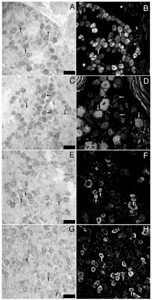
In situ hybridization for NKCC1 mRNA combined with immunohistochemistry for sensory neuron population markers in DRG: representative 40× photomicrographs of NKCC1 mRNA (A, C, E and G) combined with immunohistochemistry for peripherin (B), N52 (D), CGRP (F) and TRPV1 (H). Pairs of images are of the same field from double-labeled sections of lumbar DRGs. Upward arrows indicate examples of neurons, wherein colocalization of marker with NKCC1 mRNA was evident. Horizontal arrows in C and D illustrate NKCC1 mRNA-positive neurons that did not contain N52 immunoreactivity. Downward arrows show the converse. Scale bars=100 μm.