
Fig. 8.
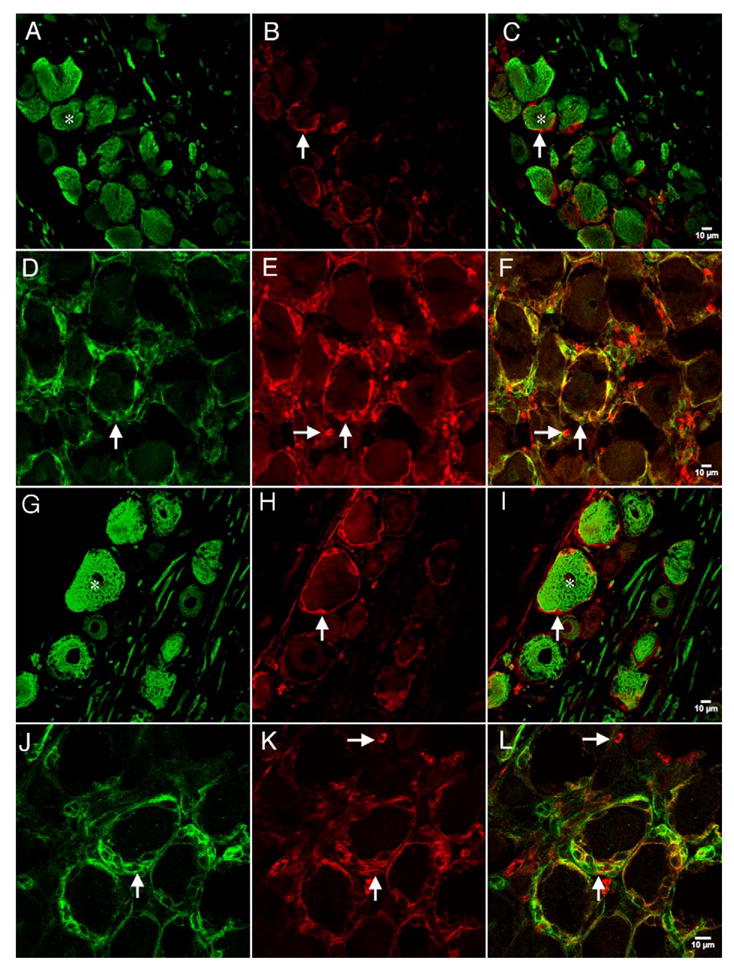
Confocal images of NKCC1 immunoreactivity with N52 or NG2 in the DRG: representative confocal photomicrographs of NKCC1 (NT) immunoreactivity utilizing the NT antibody (B and E) with N52 (A) or NG2 (D). NKCC1 (TEFS-2) immunoreactivity utilizing the TEFS-2 antibody (H and K) with N52 (G) or NG2 (J). Respective merged images are shown in panels C, F, I and L. Vertical arrows illustrate NKCC1 (NT)-immunoreactive SGCs surrounding neurons in B and C and E and F or NKCC1 (TEFS-2)-immunoreactive SGCs surrounding neurons in H and I and K and L. Vertical arrows in D and J illustrate NG2 immunoreactivity in these SGCs. Horizontal arrows in E and F and K and L demonstrate NKCC1 (NT)-immunoreactive non-neuronal cells that do not express NG2. Stars in A and C and G and I illustrate N52-immunoreactive neurons that are surrounded by NKCC1 (NT or TEFS-2, respectively)-immunoreactive cells.