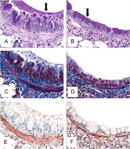
Figure 5.
Periodic acid–Schiff (A and B), Masson trichrome (C and D), and α-smooth muscle actin (E and F) staining of whole lung sections from mice challenged with OVA for 16 wk. The photomicrographs are representative of lungs removed from OVA-challenged controls (A, C, E) and FPX-treated, OVA-challenged mice (B, D, F). Goblet cell hyperplasia and mucus plugs in control mice were observed around the airways (arrow in A), but goblet cell hyperplasia was decreased and mucus plugs not evident in mice treated with 30 μg FPX (arrow in B). Enhanced collagen deposition (C, dark-blue areas) and smooth muscle cell hyperplasia (E, brown areas) were observed in control mice relative to FPX-treated, OVA-challenged mice (D and F, respectively). Original magnification: ×400.

