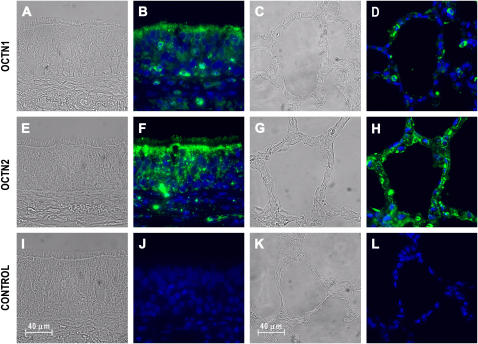
Figure 2.
Immunofluorescence analysis reveals polarized expression of the pH-dependent organic cation transporter OCTN1 and OCTN2 in human airways. After autofluorescence reduction and antigen retrieval (see Materials and Methods), human trachea and lung parenchyma sections were incubated with 5 μg/ml affinity-purified goat IgG against human OCTN1 (A–D) and OCTN2 (E–H), or nonimmune goat IgG as a negative control (I–L) followed by AlexaFluor 488–coupled rabbit anti-goat IgG. Sections were mounted in a reagent containing DAPI to stain nuclei (blue). OCTN1 (green) is primarily localized to the apical surface of tracheal epithelia (B), whereas less staining is seen in alveolar epithelia (D). Inflammatory cells in the trachea and the alveoli reveal OCTN1 immunoreactivity. OCTN2 (green) is also localized to the apical surface of airway epithelial cells (F). OCTN2 immunoreactivity is largely associated with the surface of the alveolar epithelia in the lung parenchyma (G). Figure shows bright field (A, C, E, G, I, K) and fluorescence images (B, D, F, H, J, L).

