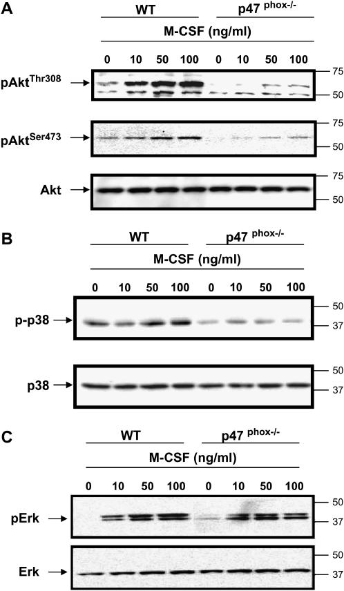
Figure 3.
Macrophages from p47phox-/- mice have decreased Akt1 and p38 MAPK activation, but not Erk activation. BMM were derived from p47phox-/- or wild-type littermate mice (WT) by growth in the presence of M-CSF (20 ng/ml) for 5 d. The cells were serum-starved overnight, then stimulated with M-CSF (10, 50, or 100 ng/ml) for 5 min or left unstimulated. (A) Akt1 activation was assessed using equal amount of protein by Western blotting with antibodies to pAktThr308 (upper panel) and pAktSer473 (middle panel). Membranes were reblotted for total Akt1 (lower panel). (B) Western blot analysis using phospho-p38 MAPK antibody and equal loading was confirmed by p38 MAPK antibody. (C) Erk phosphorylation was detected using pErk 42/44 antibody. Equal loading is shown using Erk2 antibody. Shown are representative data from macrophages obtained from six independent mice.

