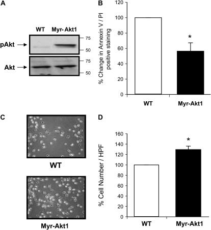
Figure 5.
Myr-Akt1–expressing macrophages have prolonged survival. BMM were generated from Myr-Akt1 or WT littermate mice by growth in M-CSF (20 ng/ml) for 5 d. The cells were serum starved for 24 h and (A) Akt1 activation was assessed by Western blotting with antibodies recognizing pAktThr308 and pAktSer473. The membranes then were reprobed for total Akt1. (B) The cells were incubated in RPMI 1640 medium for an additional 2 d to evaluate cell survival. BMM (5 × 10⁵/condition) were removed from the culture plates by Accutase, then stained with Annexin V/PI and analyzed by flow cytometry. Data shown represent macrophages from six Myr-Akt and six wild-type mice (*P < 0.05 when comparing Myr-Akt1 cell numbers versus wild-type cells). (C) The pictures were taken using ×40 objective from Olympus IX50 inverted microscope equipped with a digital camera. (D) Cells were counted and there was a significant increase in the Myr-Akt1 mice BMM (*P < 0.05 compared with the WT control).

