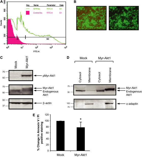
Figure 6.
Regulation of Akt1 in primary human monocytes influences cellular survival. Monocytes (10 × 106/condition) were isolated from buffy coat and transiently transfected with cDNA expressing eGFP or Myr-Akt1 using the Amaxa nucleofection. After washing, cells were plated in RPMI medium supplemented with 10% FBS and polymyxin B (10 μg/ml) and incubated at 37°C in a 5% CO2 incubator for 7 h. The monocytes were analyzed by (A) flow cytometry for transfection efficiency and (B) photography using Olympus 1X50 inverted fluorescence microscope. Images shown are phase contrast, brightfield (left panel), and fluorescence alone (right panel). (C) Cells were lysed 7 h after transfection and subjected to Western blot analysis with phospho-Akt1 antibody (upper panel). The membrane was subsequently blotted with total Akt1 antibody (middle panel) and then blotted with anti–β-actin antibody to ensure equal loading (lower panel). (D) After transfection, membrane and cytosolic fraction of the cells were also analyzed by Western blotting with anti-Akt1 antibody to show expression of Myr-Akt1 protein (upper panel). The same membranes were reprobed with the membrane-specific anti-α adaptin antibody to show the purity of the membrane fraction (lower panel). (E) Cells were stained with Annexin V/PI and analyzed by flow cytometry. Data are expressed as the mean ± SEM from six independent donors. (*P < 0.05 compared with the mock transfected samples).

