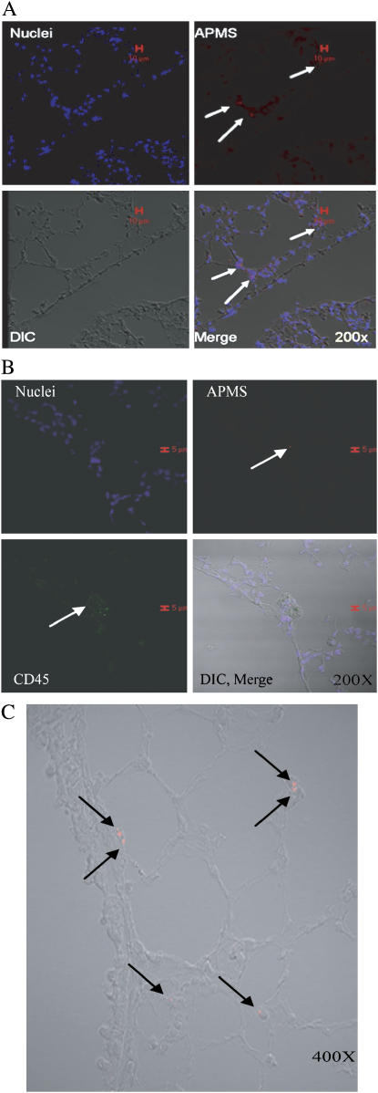
Figure 8.
APMS-TEG are found in tissues after intrapleural injection and intranasal administration. After intrapleural injection, APMS labeled with Alexa 568 (red) were found in lung tissue after 3 d (A), and occasionally in CD45+ cells in the lung after 3 d (green) (B). Cell nuclei are indicated in blue (TOTO 3). When mice nasally inhaled APMS-TEG, APMS were found in cells of the lung at 24 h (C). In all experiments, ∼ 3.3 × 10⁷ APMS were injected intrapleurally or inhaled intranasally. Red scale bars in A and B indicate 10 and 15 μm, respectively.

