Figure 5.
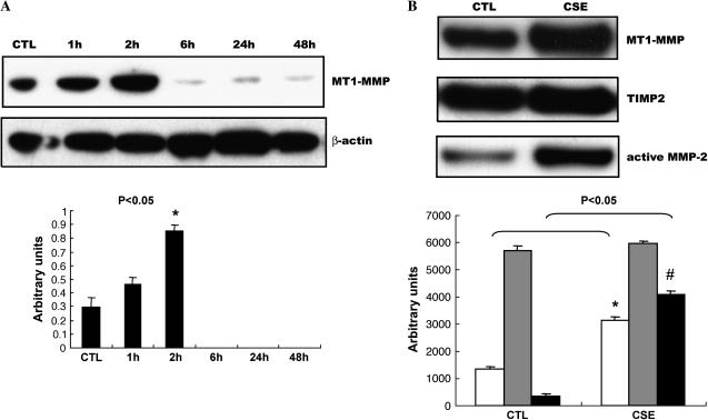
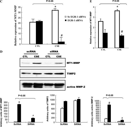
EGR-1 mediated CSE-induced MT1-MMP production. (A) NL9 cells were exposed to 20% CSE and the cell lysates were subjected to Western blot to examine MT1-MMP expression. The same membrane was probed with an anti–β-actin antibody to assess equal loading of the gel. (B) Accumulation of MT1-MMP (open bars), TIMP2 (shaded bars), and active MMP-2 (solid bars) in concentrated media of NL9 cells after 2 d CSE exposure. The EGR-1 siRNA suppress CSE-induced MT1-MMP mRNA expression (C) and protein production (D). (Panel C: solid bars, EGR-1 siRNA; open bars, scEGR-1 siRNA.) Transfected NL9 cells treated with 20% CSE for 4 h were used for mRNA assay. The concentrated media of 2-d-CSE–treated transfected NL9 cells were used for MT1-MMP, TIMP2, and active MMP-2 assay. Quantitation of Western blot immunoblots were performed by densitometric scanning of blots (A, B, and D, bottom). (Bottom of panel D: open bars, CTL; solid bars, CSE.) (E) MT1-MMP mRNA levels in mouse lung Egr-1−/− (solid bars) and control Egr-1+/+ (open bars) fibroblasts after 4 h of 5% CSE treatment.


