Figure 1.
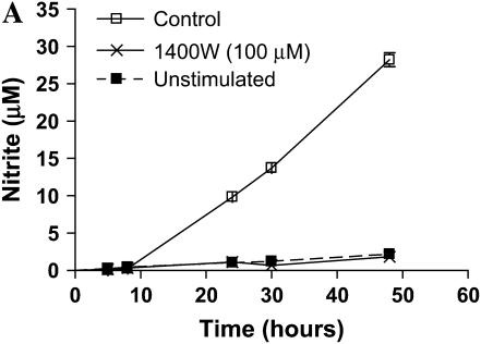
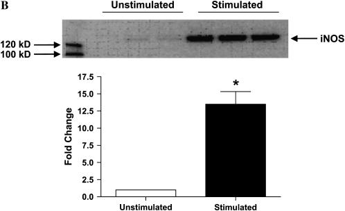
Nitrite accumulation and iNOS protein expression in stimulated LA-4 cells. LA-4 cells were exposed to LPS (10 μg/ml), IFN-γ (20 ng/ml), and TNF-α (10 ng/ml) in the presence or absence of the iNOS-specific inhibitor 1400W (100 μM) for up to 48 h. (A) Time course of nitrite production after stimulation. Cell supernatant samples were assayed for nitrite content at the specified time points (n = 3). (B) Protein extracts (25 μg), prepared from LA-4 cell lysates after 24 h stimulation with LPS (10 μg/ml), IFN-γ (20 ng/ml), and TNF-α were separated on a 4–12% denaturing polyacrylamide gel, electrophoretically transferred to PVDF membrane, and analyzed using a specific antiserum raised against iNOS and β-actin proteins. B is a representative blot of lysates from unstimulated (n = 3) and stimulated (n = 3) cells. The graph below indicates the fold change in densitometric values of iNOS protein (normalized to β-actin levels) in cells stimulated for 24 h over unstimulated controls (n = 5). *P < 0.05 versus control.


