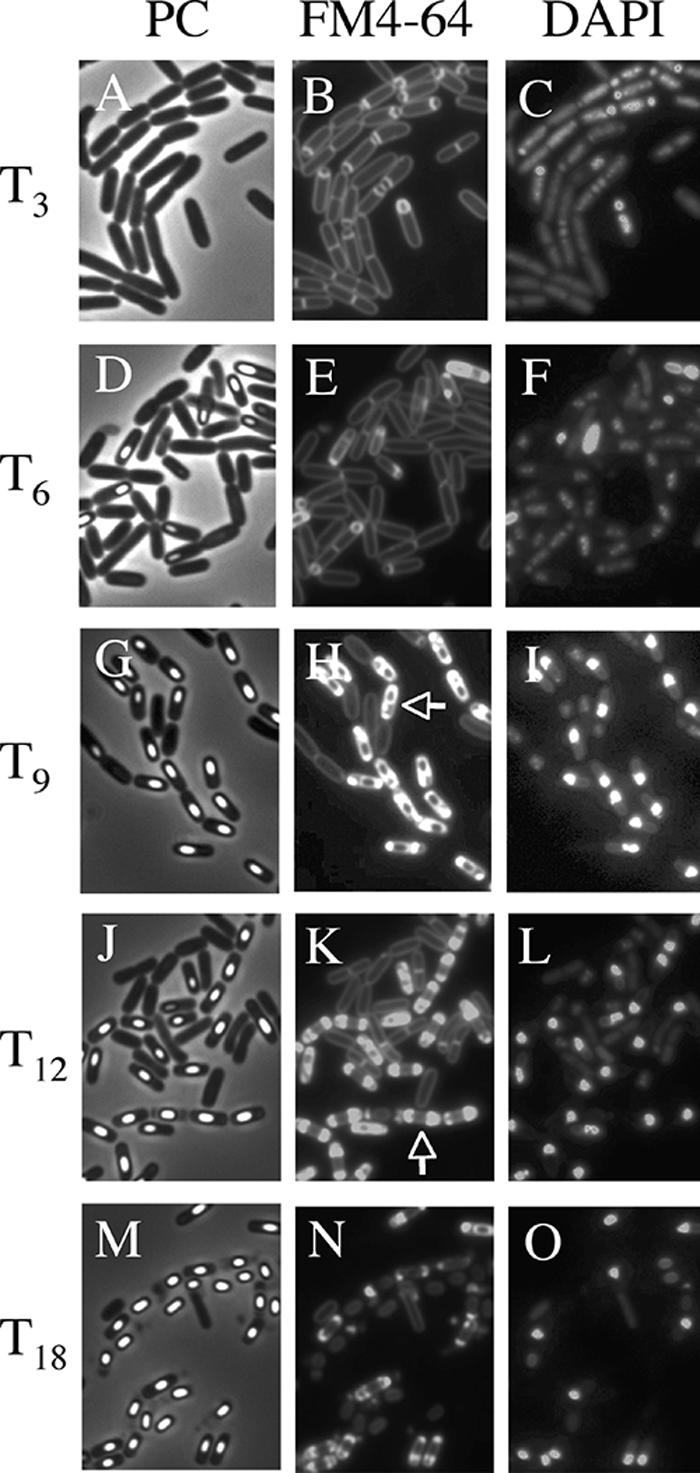
FIG. 1.

Images of mother cell in cells from liquid culture at the late stages of sporulation. Cells from strain 168 were grown to T3 (A, B, and C), T6 (D, E, and F), T9 (G, H, and I), T12 (J, K, and L), and T18 (M, N, O) in resuspension medium (8, 14). Panels A, D, G, J, and M show phase-contrast (PC) images; panels B, E, H, K, and N and panels C, F, I, L, and O show the corresponding FM4-64 staining and DAPI staining images, respectively. DAPI fails to stain the developed forespore (bright spore) DNA, but it can stain the mother cell DNA. Representative cells with intense fluorescence on one side (rupture I) (H) and both sides (rupture II) (K) are indicated with arrows.
