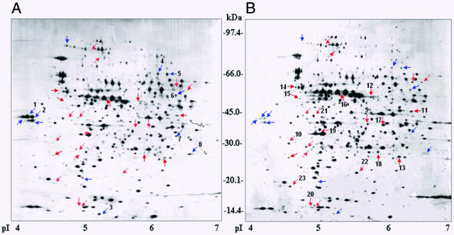
FIG. 4.
Comparative 2-D analysis of whole-cell lysates from S. enterica serovar Typhimurium UK1 wild-type (A) and UK1 ΔcadC (B) strains. Strains were adapted to acid for 1 h in NCE glucose medium (pH 4.4) containing 10 mM lysine. Protein (50 μg) was separated by IEF (pI 4 to 7) and gradient SDS-polyacrylamide gel electrophoresis (8 to 16%). Blue and red arrows indicate putative CadC-induced and -repressed proteins, respectively.

