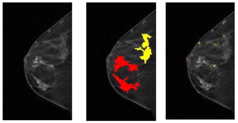
Fig 4. A 51-year-old female imaged on GE LX system. Patient had three areas identified on magnetic resonance as suspicious. All three were found to be infiltrating lobular carcinoma on final pathology. Only the subareolar mass was noted on mammography. Although all three regions with lobular carcinoma showed suspicious morphology, there are limited kinetic indications of malignancy.

(a) First postcontrast T1 image, with a DICOM stamp showing a time of 202 seconds after precontrast image.
(b) Figure 4a overlaid with map showing positive morphologic blooming (red) and near-positive morphologic blooming (yellow).
(c) Figure 4a with a kinetic map overlay showing no significant areas of washout (red) plateau (yellow).
