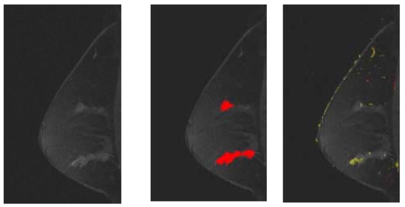
Fig 5. A 53-year-old female imaged on Siemens Magnetom system. Patient had two areas identified on magnetic resonance as suspicious. Both areas were found to be ductal carcinoma in situ Grades II-III on biopsy. The two regions identified as having suspicious morphologies match the regions with positive biopsies. Only the inferior lesion had suspicious kinetics.

(a) First postcontrast T1 image, with a DICOM stamp showing a time of 904 seconds after precontrast image.
(b) Figure 5a overlaid with map showing suspicious morphologic blooming.
(c) Figure 5a with a kinetic map overlay showing no significant areas of washout (red) and a very small area of plateau (yellow) in anterior-inferior region.
