
Figure 1. Detection of cells infected with GFP-MCMV by flow cytometry.
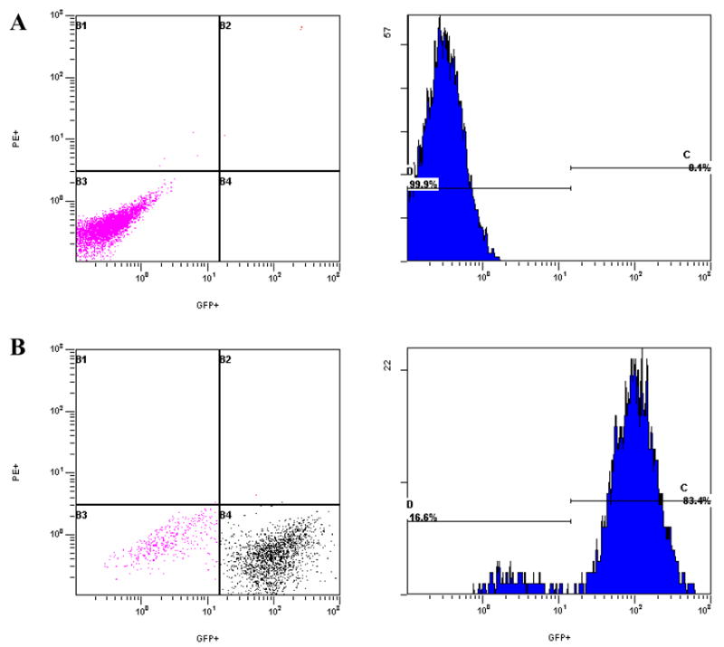
Histograms and dot plots illustrate green fluorescent protein (GFP) detection in uninfected fibroblasts and fibroblasts infected with GFP-labeled murine cytomegalovirus (GFP-MCMV). A. Uninfected NIH-3T3 fibroblasts. B. GFP-MCMV-infected NIH-3T3 fibroblasts.