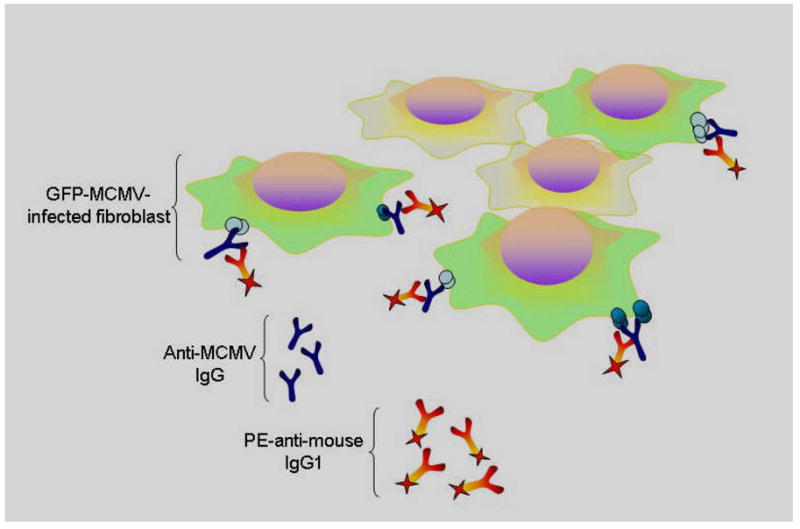
Figure 2. Schematic of flow cytometry technique to measure murine antibody responses to MCMV.

Cells infected with green fluorescent protein labeled murine cytomegalovirus (GFP-MCMV), shown as green above, are detectable by flow cytometry. These GFP-MCMV infected target cells are incubated with sera from previously infected mice, which contains anti-MCMV antibody, along with phycoerythrein labeled anti-murine IgG1 antibody. Cells that co-localize these fluorophores are detectable by dual color flow cytometry, providing a method to detect development anti-MCMV antibodies in mice.
