
Figure 4. Target cells analyzed 24 or 48 hr after infection show identical antibody binding profiles.
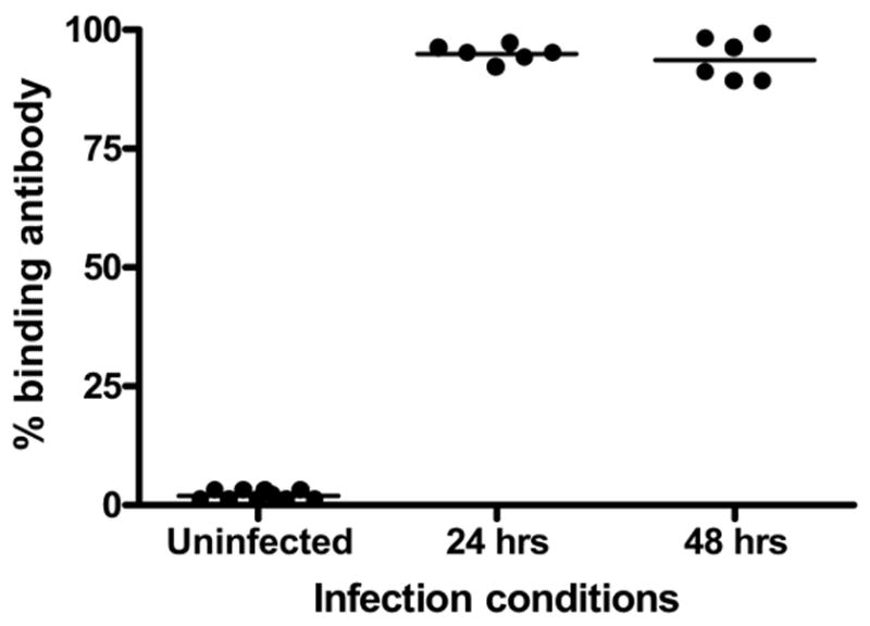
NIH-3T3 cells exposed to green fluorescent protein (GFP) labeled murine cytomegalovirus (MCMV) for 24 or 48 hours were evaluated as targets for antibody binding using MCMV-reactive standard sera and phycoerythrein (PE)-labeled anti-murine IgG1 secondary. Antibody binding is expressed as percentage of GFP positive cells which co-localized PE.