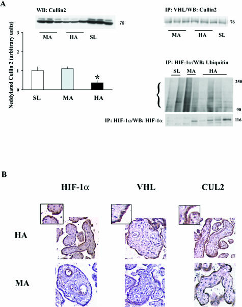
Figure 2.
VHLCBC complex interactions and immunolocalization of HIF-related molecules in placenta at different altitudes. A: Left panel, top, Western blot for Cullin-2 protein, detected at 76 kd, and neddylated Cullin-2 protein. Bottom: Densitometric analysis of neddylated Cullin-2, identified at 84 kd in Western blot (top). Right panel, top: Interaction between VHL and Cullin2 identified by immunoprecipitation with VHL followed by immunoblotting with Cullin-2. Bottom: Immunoprecipitation of HIF-1α followed by Western blotting with ubiquitin or HIF-1α. B: Immunostaining for HIF-1α, VHL, and Cullin-2. Brown color depicts positive immunoreactivity. The insets show greater magnification of the areas of interest. *P < 0.05.

