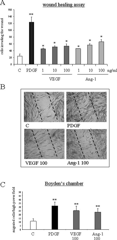
Figure 2.
VEGF and Ang-1 stimulate nonoriented migration and chemotaxis of human HSC/MFs. Nonoriented migration was assessed by means of WHA (A, B), whereas chemotaxis was assessed by means of Boyden’s chamber (C). WHA was performed on cells seeded on 24-well plates coated with collagen type I, grown to confluence in complete medium, and then incubated for an additional 24 hours in serum-free medium. An artificial lesion was generated in the cell layer to remove a linear area of cells, and then cultured cells were allowed 18 hours to migrate in the absence (control) or in the presence of increasing concentrations of human recombinant VEGF or Ang-1 or in the presence of human recombinant PDGF-BB (10 ng/ml) used as a positive control. Chemotaxis assay (C) was performed in trypsinized 24-hour-starved cells placed in Boyden’s chambers and then exposed to VEGF or Ang-1 (both used at 100 ng/ml) or to PDGF-BB (10 ng/ml) used as positive control. Data in bar graphs (A, C) represent mean ± SEM (n = 5, in triplicate) and are expressed as number of cells migrated in the artificial lesion (WHA, A) or in the filter (C), respectively. *P < 0.05 and **P < 0.01 versus control values. Representative images of nonoriented migration of human HSC/MFs under the different experimental conditions indicated are provided in B. Original magnifications, ×100.

