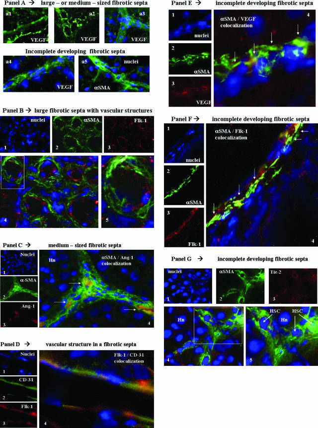
Figure 8.
In vivo localization of HSC/MFs, proangiogenic cytokines, and related receptors in fibrotic rat livers. Immunofluorescence was performed on liver cryostat sections from the liver of chronically injured rats obtained at 9 weeks in the CCl4-dependent chronic protocol for fibrosis induction. A: Reported images (green fluorescence) of immunostaining for VEGF (a1 to a4) or α-SMA (a5) in the presence or absence of concomitant nuclear stain (blue fluorescence, DAPI staining). In B, C, E, F, and G are reported the following: 1) tiny images representing image acquisition of single fluorescence always identifying in the same section nuclei (1, blue fluorescence, DAPI staining), α-SMA (2, green fluorescence), or the growth factor or receptor under analysis (3, red fluorescence; B: Flk-1; C: Ang-1; E: VEGF; F: Flk-1; G: Tie-2); 2) a larger image offering combined immunofluorescence (4, electronic merging of images); and 3) a higher digital magnification of the boxed area (5, when present). In D are reported the following: tiny images representing (see before) in the same section nuclei (1), CD-31 (2, green fluorescence), and Flk-1 (3, red fluorescence); and a larger image (4, electronic merging of images) offering combined immunofluorescence. Areas of co-localization between α-SMA and the antigen under analysis (yellow/orange fluorescence) are indicated by arrows. Hn, hepatocyte nucleus; EC, endothelial cell. Original magnifications: ×200 (Aa1–Aa3); ×400 (Aa4, Aa5, B, C, F); ×1000 (D, E, G).

