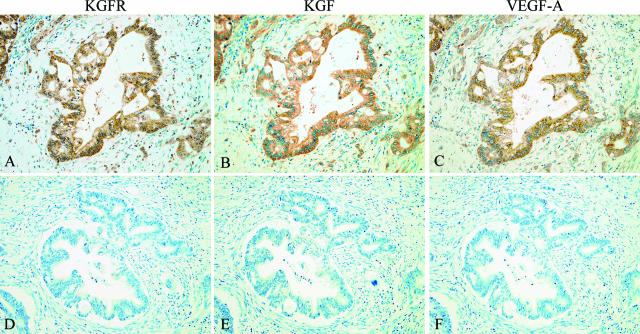
Figure 2.
Immunohistochemical analyses of KGFR, KGF, and VEGF-A in human pancreatic cancer using serial tissue sections. A–C: Characteristic staining patterns of KGFR, KGF, and VEGF-A in human pancreatic cancer cases. A: KGFR immunoreactivity was detected in the cytoplasm and cell membrane of cancer cells. B: KGF immunoreactivity was detected in the cytoplasm of cancer cells and stromal fibroblasts. C: VEGF-A immunoreactivity was detected in the cytoplasm of cancer cells and stromal fibroblasts. D–F: KGFR-, KGF-, and VEGF-A-negative cases. Immunohistochemistry, KGFR (A and D), KGF (B and E), and VEGF-A (C and F); original magnification, ×200.

