Figure 1.
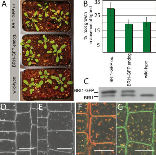
Endogenously expressed BRI1-GFP localizes to endosomes. (A) Representative pictures of rosette stage Arabidopsis grown under identical conditions. The BRI1-GFP line expressing at endogenous levels (endog.) is indistinguishable from wild type, whereas the overexpressing line (ox.) shows the reported overexpression phenotypes of narrow leaf blades and elongated, twisting petioles (leaf stalks). (B) Roots were depleted of endogenous BRs by growth on 5 μM brassinazole for 3 d. Primary root growth was assessed after three more days (n = 10 per line). Percent growth relative to untreated control is shown. The line expressing BRI1-GFP at endogenous levels is inhibited to wild-type levels, whereas the overexpressing line (ox.) shows less inhibition of root growth in the absence of ligand. (C) Immunoblot of the same lines detected with α-BRI1 antibodies. Intensity of the BRI1-GFP (top) band is much stronger than the band of endogenous BRI1 (bottom) in the overexpressing line, but both bands show the same intensity in middle lane. (D,E) Subcellular localization and levels of BRI1-GFP in root meristem epidermal cells. (D) BRI1-GFP-overexpressing line. (E) BRI1-GFP endogenous expresser. (F) BRI1-GFP (green) partially colocalizes with the endocytic tracer FM4-64 (red) after 5–10 min uptake. (G) BRI1-GFP (green) also colocalizes partially with VHA-a1-RFP (red). See Supplementary Figure 1 for individual channels of overlays. Bars, 10 μm.

