Figure 3.
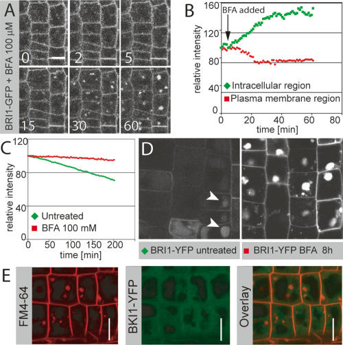
BFA increases endosomal localization of BRI1. (A) Time-lapse analysis of BFA-induced shift of BRI1 from the plasma membrane to endosomal compartments. (B) Quantification of time lapse in A. BFA induces a rapid drop in plasma membrane signal with a concomitant increase in intracellular fluorescence. A new steady state is apparently reached after ∼30 min. (C) Quantification of the effect of BFA on BRI1-YFP degradation in a pulse-chase experiment. BFA leads to a nearly complete block of BRI1 turnover. (D) BRI1-YFP localization in seedling root meristems 8 h after heat-shock-induced peak expression, with (left) or without (right) BFA treatment, taken under identical settings. BFA leads to strong accumulation of BRI1-YFP in endosomal aggregates, whereas only a weak and predominantly vacuolar BRI1-YFP signal is observed in the untreated control (arrowheads). (E) FM4-64 (red, left) and BKI1-YFP (green, middle) after 50 μM BFA treatment for 30 min. (Right) Overlay. Note the absence of BKI1 signal in BFA compartments. Bars: A,D,E, 10 μm.

