Figure 2.
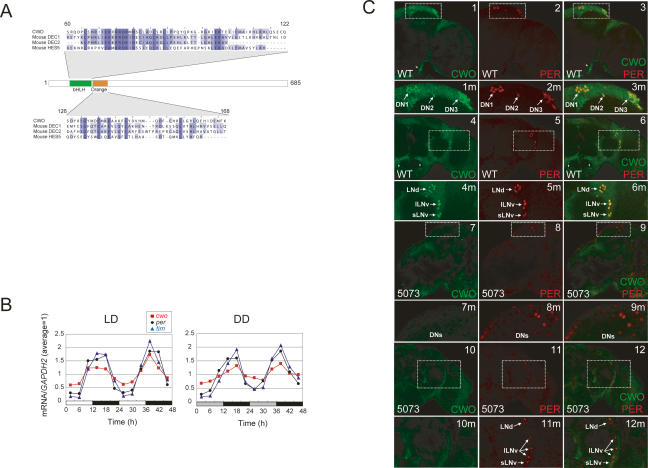
Temporal and spatial expression pattern of CWO protein. (A) cwo encodes a bHLH-ORANGE family protein. The CWO protein, 685 amino acids in length, is multiply aligned with mouse DEC1, DEC2, and HES5 proteins. Conserved amino acids in bHLH (green) and ORANGE (orange) domains are marked by blue and gaps are represented as “−”. (B) Temporal expression profiles of cwo (red square), per (black circle), and tim (blue triangle) mRNA in wild-type flies under LD and DD. Relative mRNA levels of the indicated genes were measured using a Q-PCR assay. GAPDH2 was used as an internal control. Data were normalized so that the average copy number (n = 2) over 12 time points is 1.0. (C) Spatial expression pattern of CWO in adult brains. (Panel 1) CWO immunostaining (green) in a 12-μm optical Z-stack through the right hemisphere of a wild-type brain. Three clusters of CWO-positive cells are detected in the dorsal brain. A magnified view of the boxed region is shown in panel 1m. Arrows denote DN1s, DN2s, and DN3s. Arrowheads denote additional CWO immunostaining. (Panel 2) PER immunostaining (red) in the same region shown in panel 1. Three clusters of PER-positive cells are detected in the dorsal brain. A magnified view of the boxed region is shown in panel 2m. Arrows denote the same cells described in panel 1m. (Panel 3) CWO and PER coimmunostaining in the same region shown in panels 1 and 2. Colocalization of CWO and PER immunofluorescence in this superimposed dual laser image is shown in yellow. A magnified view of the boxed region is shown in panel 3m. Arrows denote the same cells described in panels 1m and 2m. Arrowheads denote CWO staining in cells not expressing PER. (Panel 4) CWO immunostaining in a 22-μm optical Z-stack through the right hemisphere of a wild-type brain. Three clusters of CWO-positive cells are detected in the lateral brain. A magnified view of the boxed region is shown in panel 4m. Arrows denote clusters of LNds, lLNvs, and sLNvs. Arrowheads denote additional CWO immunostaining. (Panel 5) PER immunostaining in the same region shown in panel 4. Three clusters of PER-positive cells are detected in the lateral brain. A magnified view of the boxed region is shown in panel 5m. Arrows denote the same cells described in panel 4m. (Panel 6) CWO and PER coimmunostaining in the same region shown in panels 4 and 5. Colocalization of CWO and PER immunofluorescence is shown in yellow. A magnified view of the boxed region is shown in panel 6m. Arrows denote the same cells described in panels 4m and 5m. Arrowheads denote CWO staining in cells not expressing PER. (Panel 7) CWO immunostaining in an 8-μm optical Z-stack through the right hemisphere of a f05073 mutant brain. No specific CWO immunostaining is detected. A magnified view of the boxed dorsal brain region is shown in panel 7m. (Panel 8) PER immunostaining in the same region shown in panel 7. PER-positive DNs are detected in the dorsal brain. A magnified view of the boxed region is shown in panel 8m. (Panel 9) CWO and PER coimmunostaining in the same region shown in panels 7 and 8. Only PER immunofluorescence in DNs is seen in this superimposed dual laser image. A magnified view of the boxed region is shown in panel 9m. (Panel 10) CWO immunostaining in a 24-μm optical Z-stack through the right hemisphere of a f05073 mutant brain. No specific CWO immunostaining is detected. A magnified view of the boxed region is shown in panel 10m. (Panel 11) PER immunostaining in the same region shown in panel 10. Three clusters of PER-positive cells are detected in the lateral brain. A magnified view of the boxed region is shown in panel 11m. Arrows denote clusters of LNds, lLNvs, and sLNvs. (Panel 12) CWO and PER coimmunostaining in the same region shown in panels 10 and 11. Only PER immunofluorescence is detected. A magnified view of the boxed region is shown in panel 6m. Arrows denote the same cells described in panel 11m.

