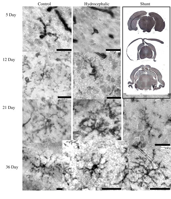
Figure 3.
Isolectin B4 antibody staining for detection of microglia (in cortical layers 2–3). Microglial morphology was observed in the cortex of in control, hydrocephalic and shunted animals. In the 5d and 12d hydrocephalic animals, a relative lack of processes on the microglia cell was evident, while the 21d and 36d hydrocephalic animals, had shorter thicker processes than control. Following shunting in both age groups, a return of fine-branched processes was seen. Scale bar = 25 μm. Low power images of brains from 36d rats at the upper right demonstrate the gross effect of shunting (lower image) on cortical thickness and ventricular volume when compared to the control (upper) and hydrocephalic brain (center).

