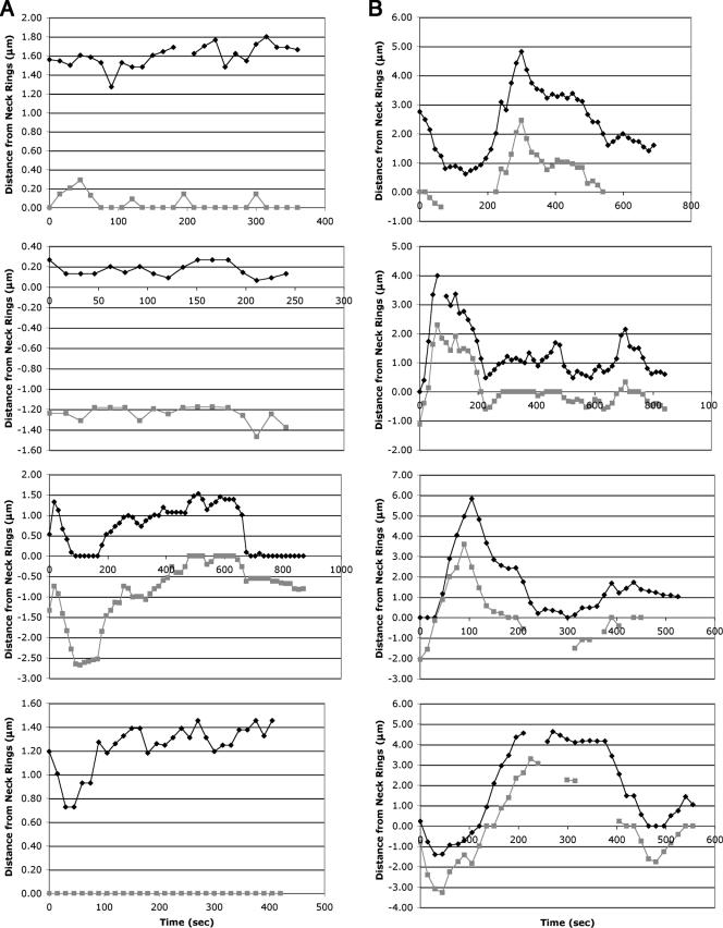
FIG. 5.
Graphical representations of SPB movements in wild-type and bud6Δ/bud6Δ diploid cells. The distances of GFP-bud6-1p spots from neck ring structures were measured at 15-s intervals in representative cells of (A) the wild type (FY23x86) or (B) bud6Δ/bud6Δ (DAY101x102) diploids carrying plasmid pDA257. Positive and negative values denote positions on the mother and daughter sides of the neck, respectively; gray and black lines distinguish the two SPBs.

