
Figure 5.
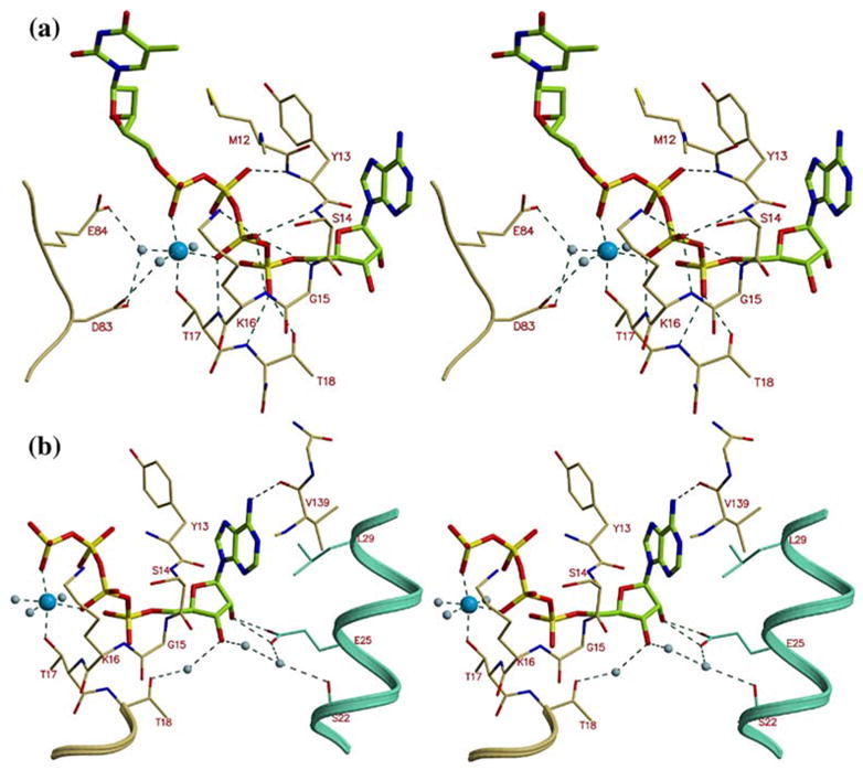
The ATP binding site in TmTK. (a) Stereo view of TP4A bound to TmTK. The phosphate groups bind to the enzyme P-loop (residues 10–16). We observe an octahedrally liganded magnesium atom. The conserved carboxylic acid residues at positions 83 and 84 interact via water molecules with the magnesium, (b) The adenosine moiety is sandwiched between two monomers that make up dimers of type II. The base is flanked by Tyr13 of one monomer and Leu29 of another. Glu25 of the neighboring subunit also directly interacts with the adenosine ribose moiety.