
Figure 2.
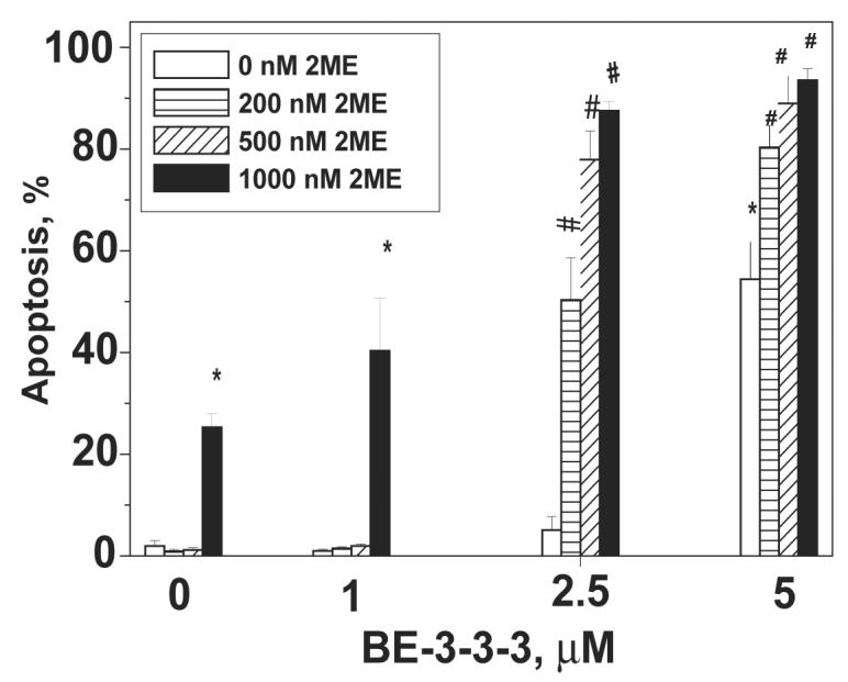
Apoptosis of MCF- 7 cells in the presence of 2ME/BE-3-3-3 combinations. Cells were treated with 2ME, BE-3-3-3, or their combinations, 24 h after plating. All of the groups also received 10 nM E2. Apoptosis was quantified using APOP-BRDU kit using flow cytometry. Data are mean ± SE from three separate experiments. * Statistically significant compared to the control (P < 0.01). #Statistically significant compared to groups treated with 2ME or BE-3-3-3 as single agents (P < 0.01).