
Figure 4.
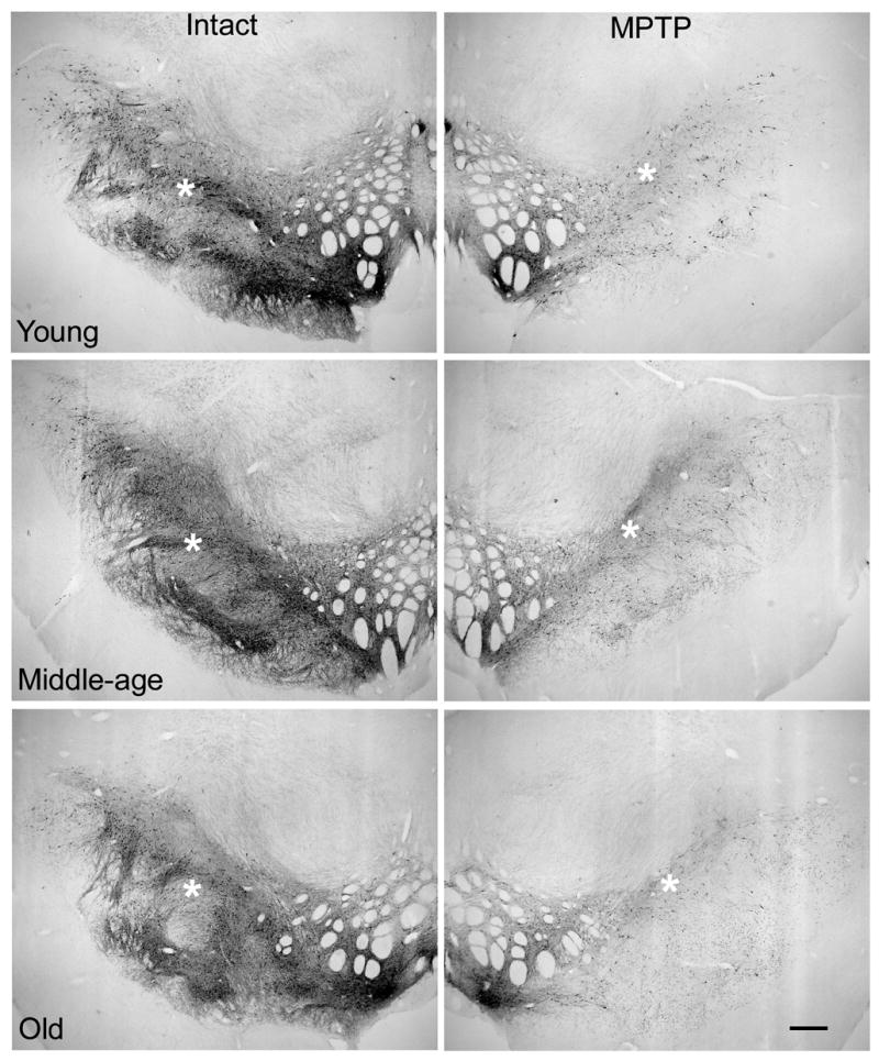
Low magnification views of TH immunoreactivity in SNC of the intact hemisphere suggest a decrease in cell number and neuropil density accompany advancing age (calibration bar=600μm). However, quantitation of THir cell numbers with stereology indicates no aging-related loss of neurons. The difference in appearance at low magnification was accounted for by a progressive increase in the frequency of THir neurons that stained lightly and decreased in cross-sectional area with advancing age (Figure 5). The expected dramatic loss of THir SNC neurons accompanied MPTP exposure in the ipsilateral hemisphere. * denote regions associated with high magnification views presented in Figure 5.