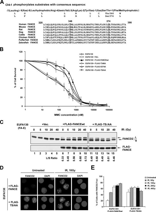
FIG. 1.
Two highly conserved Chk1 phosphorylation sites on FANCE. (A) Alignment of sequences surrounding the phosphorylation motif for Chk1 [R-X-X-(S/T)] in human FANCE with FANCE sequences in other organisms. (B) Complementation of MMC sensitivity of an FA-E lymphoblast cell line, EUFA130, with empty vector (pMMP), pMMP-FLAG-FANCE, pMMP-FLAG-T346A, pMMP-FLAG-S374A, and pMMP- FLAG-TS/AA. The indicated retroviral supernatants were generated and used to transduce EUFA130 cells. Puromycin-resistant cells were selected, and MMC sensitivity was determined as described in Materials and Methods. The values shown are the means ± standard deviations from four separate experiments. (C) Restoration of monoubiquitination of FANCD2. The indicated stably transduced FA-E lymphoblast cell lines were either untreated or exposed to IR at different doses, as indicated, and harvested after 6 h. Western blotting was performed with anti-FANCD2 or anti-FLAG antibodies. (D) Restoration of FANCD2 nuclear foci formation. EUFA130 lymphoblasts stably expressing FLAG-FANCEwt (EUFA130+FLAG-FANCE) and the double mutant FLAG-FANCE(TS/AA) (EUFA130+FLAG-TS/AA) were either untreated or treated with IR (10 Gy) and fixed 6 h later; immunofluorescence was determined using anti-FANCD2 (FI-17) antibody. Magnification, ×630. (E) Quantification of FANCD2 foci. Cells with more than four distinct foci were counted as positive; 200 cells/sample were analyzed. The values shown are the means ± standard deviations from three separate experiments. Vec, empty vector.

