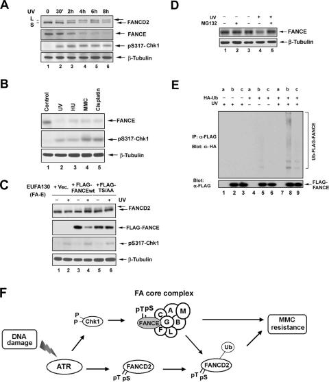
FIG. 6.
FANCE phosphorylation by Chk1 promotes its degradation. (A) HeLa cells were either untreated or treated with UV irradiation at 60 J/m2 and incubated for different periods of time as indicated before lysis. Whole-cell extracts were immunoblotted with the indicated antibodies. An anti-β-tubulin blot was used as a loading control. (B) HeLa cells were synchronized by a double-thymidine block and then released into S phase. One hour after release, the cells were either untreated (Control) or treated with UV (60 J/m2, 15 h), HU (2 mM, 24 h), MMC (160 ng/ml, 24 h), or cisplatin, (10 μM, 24 h). Whole-cell extracts were analyzed by Western blotting with the indicated antibodies. An anti-β-tubulin blot was used as a loading control. (C) EUFA130 lymphoblasts were stably expressed with pMMP (empty vector [Vec]), FLAG-FANCEwt, or FLAG-FANCE(TS/AA) (the double mutant) as indicated. Cells were either untreated or treated with UV (60 J/m2); after 8 h, whole-cell extracts were analyzed by Western blotting with the indicated antibodies. An anti-β-tubulin blot was used as a loading control. (D) U2OS cells were either untreated or treated with UV (60 J/m2) and incubated for 3 h with or without the addition of 25 μM MG132 to the indicated samples during the final 2 h in cell culture. Whole-cell extracts were analyzed by Western blotting with the indicated antibodies. An anti-β-tubulin blot was used as a loading control. (E) FANCE ubiquitination in vivo. U2OS stably expressing empty vector (a), FLAG-FANCEwt (b), or and FLAG-FANCE(TS/AA) (c) were transiently transfected without or with a cDNA encoding HA-Ub; after 48 h of transfection, cells were untreated or treated with UV (60 J/m2) and incubated for 2 h before cells were lysed in SDS denaturation buffer. FLAG-FANCEwt and the double mutant protein were isolated by anti-FLAG antibody immunoprecipitation. Immune complexes were run on SDS-PAGE gels and immunoblotted with anti-HA or anti-FLAG antibodies. (F) Schematic model showing the activation of the FA/BRCA pathway by the ATR-Chk1 pathway. DNA damage or replication arrest (MMC, UV, IR, or HU) activates the ATR-dependent phosphorylation of FANCD2 (6) and the Chk1-dependent phosphorylation of FANCE. Both monoubiquitinated FANCD2 and phosphorylated FANCE are required for MMC resistance. A nonubiquitinated mutant of FANCD2 (K561R) or the nonphosphorylated mutant FANCE(TS/AA) fails to correct MMC hypersensitivity.

