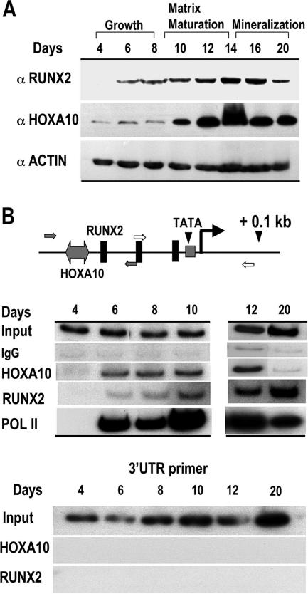
FIG. 6.
HOXA10 protein and recruitment to the Runx2 promoter in primary calvarial cells during osteoblast growth and differentiation. (A) Western blot analyses for RUNX2 and HOXA10 protein expression are shown during stages of growth and differentiation of isolated primary calvarial osteoblasts as indicated (antibody information is provided in Materials and Methods). Protein profiles of actin demonstrate equivalent amounts of total protein loaded in the gel. (B) ChIP studies of the Runx2 proximal promoter locus during growth and differentiation. The top panel illustrates the positions of regulatory elements and primers used to amplify the Runx2 promoter-specific DNA fragments. Open arrow, RNA Pol II; solid arrow, HOXA10 and RUNX2 occupancy. For the middle panel, ChIP analysis was performed on the indicated days with antibodies for HOXA10, RUNX2, or RNA Pol II (see Materials and Methods). One percent of the soluble chromatin fraction was taken as the input fraction. IgG is shown as an antibody control. The lower panel shows the control for ChIP primer specificity.

