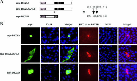
FIG. 3.
Subcellular localization of BSX1AΔNLS. (A) Schematic representation of myc-tagged proteins expressed in Hs683 cells. HD, homeodomain. (B) Immunocytological analyses of myc-tagged protein-transfected Hs683cells. The images on the left (green) are myc-immunoreactive signals, and those on the right show BSX1A -or BSX1B-immunoreactive cells (red). Note that BSX1A was detected in the nuclei and BSX1AΔNLS and BSX1B in the cytoplasm.

