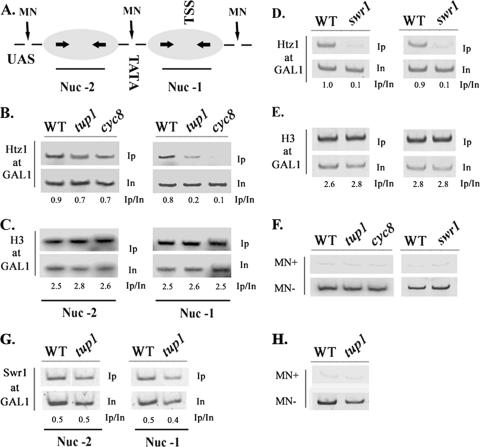
FIG. 6.
Tup1-dependent Htz1 deposition involves only the GAL1-proximal nucleosome. (A) Schematic representation of the GAL1 promoter nucleosomal organization. The positions of the distal (Nuc −2) and proximal (Nuc −1) nucleosomes relative to the UASGAL, TATA element, and TSS are indicated along with MNase-sensitive sites. Arrows within nucleosomes represent the positions of primers used for PCR amplification. (B) Histone Htz1 and histone H3 (C) depositions at the distal (Nuc −2) and proximal (Nuc −1) GAL1 nucleosomes were monitored in glucose grown wild-type (WT), tup1, and cyc8 strains following chromatin IPs and PCR amplification using nucleosome-specific primers. Histone Htz1 (D) and histone H3 (E) deposition at nucleosome −2 and nucleosome −1 was monitored in wild-type and swr1 strains by chromatin IPs and PCR amplification as described above. Numbers indicate immunoprecipitated/input ratios. (F) The extent of MNase digestion in the above-described experiments was confirmed by PCR amplification of MNase-digested (MN+) or untreated (MN−) input DNA obtained from the indicated strains using primers spanning the region of both nucleosome −2 and nucleosome −1. (G) Swr1 recruitment on either nucleosome (nucleosome −2 and nucleosome −1) was monitored by chromatin IP in WT and tup1 strains grown in glucose as described above (B). (H) MNase treatment was confirmed as described above (F).

