FIG. 1.
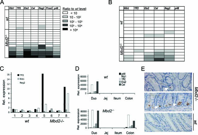
Expression of ExPa genes in wild-type and Mbd2−/− mice. (A) Real-time quantitative RT-PCR analysis of ExPa gene expression in the colon of nine wild-type and 13 Mbd2-null BALB/c mice. Each horizontal row corresponds to one mouse. Values represent the fold overexpression of genes normalized to average expression in wild-type samples. (B) The partial penetrance of ExPa gene overexpression is independent of the mouse strain. Detection of ExPa gene expression levels in the colon of eight wild-type and eight Mbd2-null C57BL/6 mice by real-time RT-PCR as in panel A. (C) ExPa gene expression levels in colons isolated from 4-day-old BALB/c wild-type and Mbd2-null mice. The expression levels were analyzed by real-time RT-PCR and were normalized to the average expression in wild-type samples. (D) Real-time RT-PCR analysis for expression patterns of ExPa genes in four intestinal segments (Duo, duodenum; Jej, jejunum) of wild-type and Mbd2-null mice. Overexpression levels are calculated compared to the levels in the wild-type colon. (E) Immunohistochemical detection of TFF2 in colonic epithelium of Mbd2-null mice (top and middle panels) and wild-type mice (bottom panel). Positively stained enterocytes (brown spots; white arrows show examples) and goblet cells are only seen in mutant crypts.

