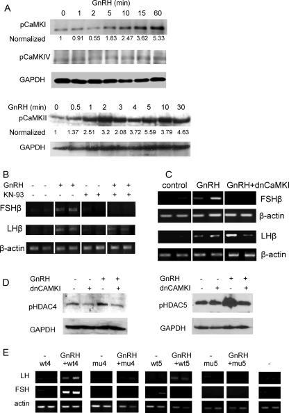
FIG. 6.
GnRH derepression of the FSHβ gene involves CaMKI. (A) Western analysis was carried out to detect phosphorylated forms of CaMKI, -II, and -IV in αT3-1 cells after exposure for 0 to 60 min to GnRH using antisera as marked; GAPDH is shown as a loading control, to which CaMK levels were normalized, and these values are expressed as a ratio to the levels in untreated cells. (B) αT3-1 cells were exposed to GnRH (10 nM for 8 h) and/or the CaMK inhibitor KN-93 (10 μM; added 30 min before the GnRH) before RT-PCR analysis of FSHβ and LHβ mRNA levels. (C) Similarly, the dnCaMKI expression vector (1.5 μg) was transfected into cells 16 h before treatment with GnRH (10 nM for 24 h), and the effect on GnRH derepression of both genes was assessed by RT-PCR in the same way. Transfections and treatments were carried out in duplicate. (D) Levels of phosphorylated HDAC4 and -5 (pHDAC4 and -5) were assessed by Western analysis in control cells and cells treated with GnRH (2 h), with or without transfection (24 h before) of the dnCaMKI construct. (E) Wild-type (wt) or mutant (mu) HDAC4 and -5 constructs (as described in the legend of Fig. 5) were overexpressed in the presence or absence of GnRH, and their effects on the derepression of LHβ and FSHβ were assessed by RT-PCR, as described in the legend of Fig. 3. Transfections and treatments were carried out in duplicate.

