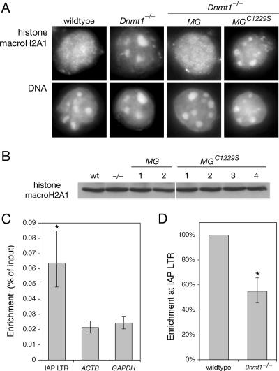
FIG. 4.
Methylation-dependent localization of histone macroH2A1. (A) Immunofluorescence with anti-histone macroH2A1 antibody. The regions of intense Hoechst DNA staining correspond to pericentric heterochromatin. Both clones of Dnmt1−/− MG and all four clones of Dnmt1−/− MGC1229S showed the same result (not shown). (B) IB of cell lysates with anti-histone macroH2A1 antibody. The lanes correspond to those shown in Fig. 1C. wt, wild type. (C) Association of histone macroH2A1 with the IAP LTR as determined by ChIP. The enrichment at the LTR is highly significant compared to the signal at ACTB and GAPDH (P = 0.001 and P = 0.002, respectively; the asterisk indicates statistical significance). Error bars represent standard deviations of the means. Quantitative real-time PCR was performed on samples from at least three independent biological replicates. (D) Methylation-dependent association of histone macroH2A1 with the IAP LTR. ChIP was performed with wild-type and Dnmt1−/− cells. The enrichment in Dnmt1−/− cells was significantly lower than that in wild-type cells (P = 0.028; the asterisk indicates statistical significance). The error bar represents the standard deviation of the mean.

