FIG. 5.
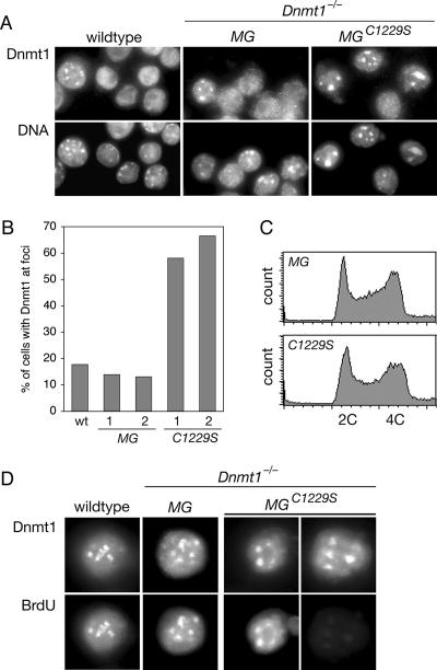
DNMT1C1229S is mislocalized to pericentric heterochromatin. (A) Immunofluorescence with anti-DNMT1 antibody. Representative fields indicate the higher fraction of Dnmt1−/− MGC1229S cells with DNMT1 foci. The regions of intense Hoechst DNA staining correspond to pericentric heterochromatin. Both clones of Dnmt1−/− MG and all four clones of Dnmt1−/− MGC1229S showed the same result (not shown). (B) The percentage of cells with DNMT1 foci was determined in wild-type (wt) cells and in cells from two clones each of Dnmt1−/− MG (“MG”) and Dnmt1−/− MGC1229S (“C1229S”). Approximately 300 to 400 cells were counted for each sample. (C) Cell cycle distribution of asynchronous culture as determined by flow cytometry. Cells were fixed in ethanol and treated with RNase A, and DNA was stained with propidium iodide. The wild type, Dnmt1−/−, both clones of Dnmt1−/− MG (“MG”), and all four clones of Dnmt1−/− MGC1229S (“C1229S”) showed the same distribution (not shown). (D) Costaining of DNMT1 and BrdU after a 15-min pulse with BrdU. All wild-type and Dnmt1−/− MG cells with DNMT1 foci were BrdU positive. In Dnmt1−/− MGC1229S cells, some cells with DNMT1 foci were BrdU positive, and some were BrdU negative.

