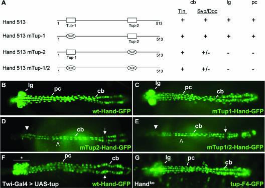
FIG. 6.
Tup is a direct transcriptional regulator of the Hand dorsal vessel enhancer. (A, left) Schematic of the Hand 513-bp HCH enhancer and three Tup site mutant versions thereof. (A, right) Function of wild-type and mutated Hand-GFP DNAs in transgenic embryos. Activities of the various enhancers in Tin or Svp/Doc cardioblasts (cb), lymph glands (lg), or pericardial cells (pc) are indicated as positive (+), negative (−), or greatly reduced (+/−). (B and C) Normal cellular expression profile of GFP in embryos harboring the wt-Hand-GFP or mTup1-Hand-GFP transgenes. (D and E) Representative embryos expressing the mTup2-Hand-GFP or mTup1/2-Hand-GFP construct. With both mutated DNAs, GFP is observed in Tin cardioblasts but absent from the lymph glands (closed arrowheads) and pericardial cells (open arrowheads). Reporter expression is greatly diminished in Svp/Doc cardioblasts (arrows) with both mutations. (F) Twi-Gal4>UAS-tup embryo expressing the wt-Hand-GFP marker. This forced expression condition results in an expanded population of prohemocytes in the lymph glands (horizontal bar and asterisk) and a disorganized heart region (arrow). (G) Expression of the tup-F4-GFP transgene in a homozygous Handko mutant embryo. tup expression appears to be normal in cardioblasts and pericardial cells but reduced in the lymph glands. All embryos are at stage 16 of development and oriented with the anterior to the left.

